
Welcome to the first installment of our new series, “Instrument of the Month”, where we’ll highlight one of the many instruments at the Molecular Imaging Center (MIC). The purpose of the series is to bring awareness to the community regarding our imaging resources. Since we currently manage 20 instruments in our facility, very few users know about all of the imaging modalities that are available at the MIC. A table listing all of our instruments and their specs can be found here.
To make things a bit more readable and less dry (hopefully), the format will be a Q & A style “interview”. Without further ado, here we go!
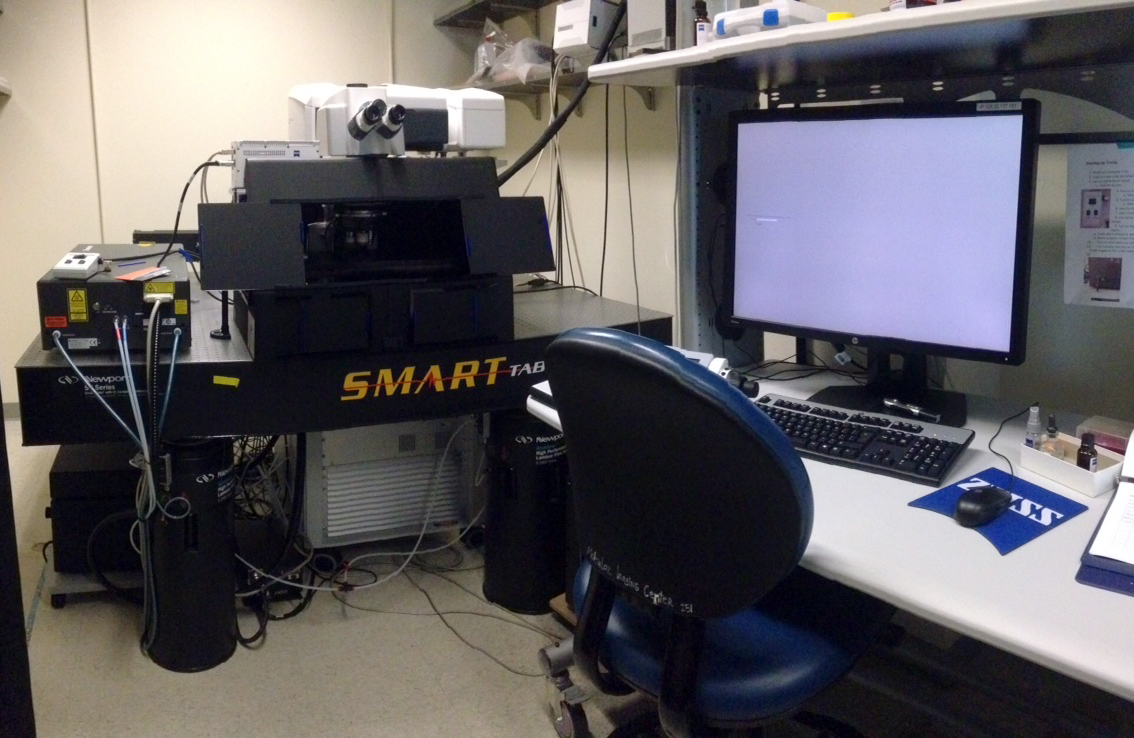
What is “Trinity”?
Trinity is a Zeiss LSM 880 with non-linear optics (NLO) for 2-photon imaging and Airyscan for super resolution (more on that later). It’s one of 3 new instruments acquired as part of the BrainMIC collaboration with Zeiss.
Why the name Trinity?
All MIC instruments have traditionally been nicknamed with sci-fi references in mind. Since we received 3 new instruments around the same time, Holly decided to name them Trinity, Neo, and Morpheus after the characters from The Matrix trilogy. We hope that they’re as powerful as their namesakes! In addition, Trinity has 3 different imaging modalities: spectral confocal, 2-photon, and Airyscan.
What laser lines does Trinity have?
– Diode 405 nm
– Argon 458, 488, 514
– DPSS 561, HeNe 594, HeNe 633
– Coherent Chameleon 690-1040 nm (2-photon imaging)
What objectives does Trinity have?
Trinity can hold 4 objectives on the main turret. Currently there is a 5X/0.25 (air), 20X/1.0 (water), 40X/1.0 (water), and 63X/1.4 (oil). We also have a 20X/0.8 (air) and a 20X/1.0 SCALE objective for cleared samples (refractive index = 1.38, working distance = 5.6mm).
Tell me about the basic features of Trinity.
Trinity is an upright laser scanning confocal with 2 PMTs, a 34-channel GaAsP spectral system, and a 2-channel Airyscan detector for 1-photon imaging. It also has a BiG.2 binary GaAsP NDD for multiphoton imaging. There’s a large motorized stage sufficient for electrophysiology studies. The black box/enclosure provides a more stable and dark environment for multiphoton imaging.
I’m already using Rachel (the LSM 780). What’s new or special about Trinity?
Trinity is like Rachel on steroids. The LSM 880 features water-cooled galvos and water-cooled detection, which means an increase in scan speed or field of view. The new BiG.2 NDD is more stable than previous versions, as it no longer has a fan in the unit. Finally, Airyscan allows for significantly improved resolution, up to 1.7-fold, using standard confocal sample prep and imaging protocols. And good news for Rachel users – we are not charging for training on Trinity if you’re already trained on the 780.
What is Airyscan?
Airyscan is a new technology developed by Zeiss, where instead of having a single PMT or a linear array of detectors (as is used for spectral imaging), there is an array of 32 detectors in a honeycomb shape. The smaller detectors allow for better resolution, while collectively, the honeycomb collects light from the entire beam path. (The fundamental principle is similar to mapping out a point spread function over the Airyscan detector.) After imaging, there is an additional post-processing step where a deconvolution algorithm is applied. The result is a higher resolution image with better signal-to-noise ratio. The AiryScan detector is also more sensitive, as it collects more light through the wider pinhole. The Airyscan detector can also be used as an additional confocal detector if you need a 4th track.
Is Airyscan super-resolution microscopy?
Yes! The system is capable of pushing the limits of resolution by 1.7-fold with green light. While it cannot achieve the 10 nm resolution of the localization methods (PALM/STORM), it does not require any changes to your protocols that are optimized for standard confocal imaging. The resolution is very similar to the SIM method, but much faster and easier for the end-user!
OK, enough theory. What do REAL Airyscan images look like, compared to confocal?
You can see some examples from Zeiss’s marketing materials on their Airyscan site, but I thought it would be cool to show you an actual image acquired on Trinity. Below are 2 images of Invitrogen Fluo prepared slide #2: BPAE cells stained with Texas Red F-actin, BODIPY FL microtubules, and DAPI. These images were taken with the 63X/1.4 oil objective (with 2X zoom). On the left is the normal confocal image (pinhole set at 1 A.U.); on the right is the Airyscan image, post-processed. Scale bar = 10 µm.

This explanation was super confusing. Where can I find more information?
Check out this very long, but extremely informative webinar on the Zeiss LSM 880 with Airyscan – embedded below. (Skip to the 6:00-minute mark to get to the segment about the 880; 17:50 is where the Airyscan section begins.)
Who do I contact for more information about the microscope, training, etc.?
You can email Jen or Holly for more information. The MIC training page lists the steps on how to get trained on all of our instruments.

[…] as part of the Zeiss/BRAIN-MIC initiative. Since there were 3 instruments, we named them Trinity, Neo, and Morpheus, after the main characters in The Matrix […]
LikeLike
[…] Here’s a little news item we wrote for the new Airyscan Fast Module, now available on Trinity. […]
LikeLike